
Last Updated May 28, 2026
Cell structure, membranes, and organelles examine how cells are physically organized, how that organization makes living function possible, and why cellular architecture is inseparable from metabolism, transport, signaling, information flow, repair, stress response, disease, ecology, and biotechnology. A cell is not a random mixture of biomolecules suspended in water. It is a bounded, compartmentalized, mechanically organized, chemically differentiated, and dynamically regulated system. Membranes create selective boundaries. Organelles create specialized environments. Cytoskeletal systems maintain shape and direct movement. Transport systems move matter, energy, and information across space. Cellular structure therefore matters because biological function depends on organization.
This article develops Cell Structure, Membranes, and Organelles as a foundational article within the Biology knowledge series. It treats cellular architecture not merely as anatomical description, but as one of biology’s major explanatory frameworks: the physical logic through which molecular processes become coordinated living systems. The arrangement of membranes, nucleus, mitochondria, chloroplasts, endoplasmic reticulum, Golgi apparatus, lysosomes, peroxisomes, vacuoles, cytoskeleton, vesicles, and membrane contact sites determines how cells regulate internal chemistry, maintain gradients, synthesize and traffic molecules, harvest energy, degrade damaged components, sense environments, and respond to stress.
Main Library
Publications
Article Map
Biology
Related Topic
Chemistry
Related Topic
Earth Science
Related Topic
Environmental Science
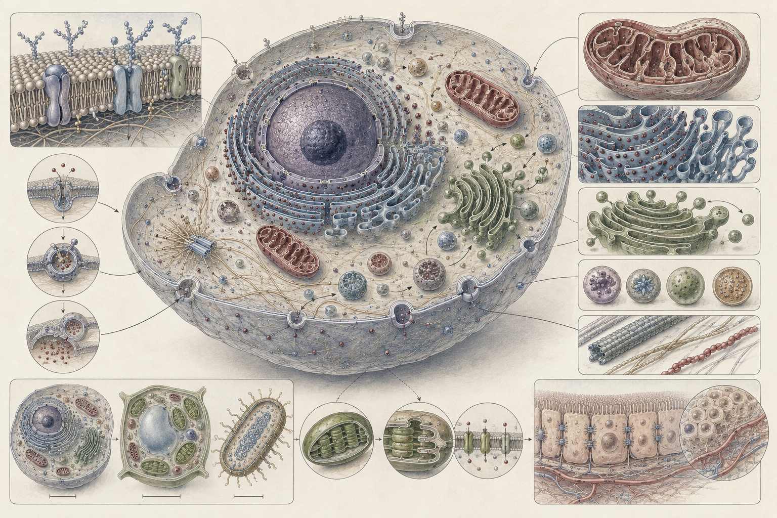
The article develops cell structure, membranes, and organelles across cellular compartmentalization, plasma membranes, membrane transport, lipid bilayers, membrane proteins, the fluid mosaic model, ion gradients, diffusion, osmosis, nucleus, chromatin organization, mitochondria, chloroplasts, endoplasmic reticulum, Golgi trafficking, lysosomes, peroxisomes, vacuoles, cytoskeleton, vesicular transport, membrane contact sites, prokaryotic and eukaryotic contrasts, ecological cell biology, marine stress biology, biomedical pathology, biotechnology, imaging, and computational cell biology.
The article also extends cell structure into quantitative and computational biology through diffusion and membrane-flux models, surface-area-to-volume scaling, compartment-volume ratios, organelle network metrics, vesicle-traffic simulations, mitochondrial capacity indices, morphometry-style measurement tables, cell-growth fitting, transport-limited uptake, compartment condition scoring, R workflows, Python workflows, SQL provenance structures, and a linked full-stack GitHub repository containing Python, R, Julia, Fortran, Rust, Go, C, C++, SQL, notebooks, data files, validation notes, and reproducibility documentation.
Why Cell Structure Matters
Biology explains living systems not only by listing their components, but by showing how those components are arranged and coordinated. Nowhere is this clearer than in the cell. A cell is not a random mixture of biomolecules suspended in fluid. It is a structured, compartmentalized, and dynamically regulated system in which membranes define boundaries, organelles create specialized environments, and transport systems move matter, energy, and information in controlled ways. Cell structure therefore matters because function depends on organization.
This principle is one of the deepest continuities between cell biology and the broader scientific understanding of life. Membrane boundaries create biological space. Compartments isolate incompatible processes while still permitting coordination. Organelles specialize biochemical work. Cytoskeletal systems maintain form while enabling movement and transport. The scientific study of cell structure is therefore also the study of how living order becomes physically organized at the smallest complete level of life.
Cell structure also matters because failure of cellular architecture often becomes failure of function. Membrane defects alter transport and signaling. Mitochondrial disruption affects energy, redox balance, and cell death. Lysosomal failure disrupts turnover. Cytoskeletal defects alter transport, division, and mechanics. Nuclear disorganization can affect gene expression and genome maintenance. The architecture of the cell is therefore causal, not decorative.
The same principle extends beyond the laboratory. Marine organisms tolerate salinity, pressure, oxygen variation, and temperature stress through cellular structures. Plants survive drought and regulate water through vacuoles, membranes, chloroplasts, and cell walls. Microbes transform soils, sediments, and wastewater through envelope structures, transport systems, and internal metabolic organization. Medicine, ecology, biotechnology, and environmental science all depend on the fact that cellular structure governs biological possibility.
The Cell as an Organized System
A cell is a bounded system in which many processes occur simultaneously without collapsing into chemical disorder. Genetic information must be stored and expressed, energy must be harvested and distributed, proteins must be synthesized and trafficked, signals must be received and interpreted, waste must be degraded or expelled, and internal composition must be regulated despite constant environmental change. This requires not merely molecular presence but spatial organization.
Cell structure makes that possible. Different reactions occur in different places, often under different chemical conditions. The existence of compartments allows cells to separate functions while preserving overall coordination. In this sense, the cell is not simply a basic unit because it is small. It is basic because it is the smallest level at which living organization is complete enough to support continuous, regulated life.
This also means that cellular structure is dynamic. Membranes remodel. Vesicles bud and fuse. Organelles divide, move, and interact. Cytoskeletal fibers assemble and disassemble. Mitochondria fuse, fragment, and relocate. The nucleus reorganizes chromatin. The cell is therefore a structured system, but not a static one. Its organization is maintained through regulated motion, exchange, and repair.
This dynamic view is especially important for modern biology. A textbook diagram can make cells appear as fixed arrangements of parts. Real cells are temporal systems. Their architecture changes with nutrient availability, cell-cycle state, stress, developmental program, infection, mechanical force, temperature, oxygen availability, and environmental chemistry. The study of cell structure is therefore also the study of cellular responsiveness through physical organization.
Membranes, Boundaries, Selectivity, and Biological Space
Membranes are among the most important structures in biology because they create the boundaries on which living organization depends. The plasma membrane separates the cell from its surroundings, while internal membranes separate organelles from the cytosol and from one another. These boundaries are not passive walls. They are selectively permeable interfaces through which cells regulate transport, sense environments, preserve gradients, and coordinate exchange.
Without membranes, there would be no stable intracellular environment, no controlled transport, no chemiosmotic energy systems, and no effective compartmentalization of biochemical work. Membranes therefore make biological space possible. They transform chemistry into organized, bounded process by distinguishing inside from outside and one internal environment from another.
Membrane selectivity is especially important because cells must regulate what enters, what exits, and what remains separated. Ions, metabolites, proteins, lipids, signaling molecules, toxins, water, and wastes do not move freely in ways that would preserve life automatically. Channels, pumps, carriers, transporters, receptors, and vesicular pathways make exchange selective and regulated. Biological membranes are therefore active interfaces, not inert barriers.
This is why membrane biology connects directly to physiology, ecology, and disease. Osmoregulation, nutrient uptake, nerve signaling, immune recognition, drug delivery, toxin response, pathogen entry, marine salinity tolerance, plant water stress, and microbial survival all depend on membrane structure and transport.
Membranes also connect cell biology to energy. Mitochondrial inner membranes and chloroplast thylakoid membranes organize electron transport and proton gradients. Bacterial membranes support respiration, transport, and signaling. Membrane architecture is therefore not only a boundary system; it is one of the key structures through which cells convert gradients into work.
The Fluid Mosaic and Membrane Function
Modern biology often describes membranes through the fluid mosaic model, which emphasizes that biological membranes are dynamic assemblies of lipids, proteins, sterols, carbohydrates, and associated molecules rather than rigid shells. Lipids provide the bilayer structure, while embedded and associated proteins support transport, signaling, adhesion, enzymatic activity, recognition, and structural anchoring. This means that membrane function depends on both composition and mobility.
The significance of this model is functional as much as structural. A membrane must be stable enough to preserve the cell’s integrity, but fluid enough to permit transport, receptor movement, vesicle fusion, and structural adjustment. Biological membranes therefore exemplify a wider principle of living order: successful structure in biology is often dynamic rather than fixed. The membrane is organized, but it is organized as a flexible interface.
Membrane fluidity is environmentally sensitive. Temperature, lipid saturation, sterol composition, salinity, pressure, oxidative stress, and chemical exposure can alter membrane behavior. This is especially relevant in marine biology, microbial ecology, plant stress biology, and disease. Organisms often adjust membrane composition to preserve function under changing conditions.
In that sense, the membrane is both a structural boundary and a responsive material system. It is one of the clearest places where biological organization, physical chemistry, and environmental constraint meet.
The fluid mosaic model also prevents an overly mechanical view of cells. Membranes are not merely bags that contain cellular contents. They are active surfaces where signals are received, gradients are preserved, transport is regulated, pathogens interact, drugs bind, vesicles traffic, and identity is displayed. The cell surface is one of the major interpretive interfaces between organism and environment.
The Nucleus, Information, Control, and Cellular Coordination
In eukaryotic cells, the nucleus houses the genome and serves as a central site of information storage, transcriptional regulation, genome maintenance, and cellular coordination. Its double membrane, nuclear pores, chromatin organization, nucleolus, lamina, and relation to the cytoskeleton all matter because genetic information must not only be stored, but made accessible and regulated under changing conditions. The nucleus is therefore not merely a container for DNA. It is part of a larger system of information control.
This matters because the cell depends on regulated gene expression rather than constant undifferentiated activity. Development, stress response, metabolism, division, differentiation, immune activation, and repair all require gene expression to be timed, localized, and modulated. The nucleus contributes to living order by making information both stable and usable, and by linking heredity to ongoing physiological function.
Nuclear organization also affects how genetic information is interpreted. Chromatin structure, nuclear domains, transcriptional neighborhoods, and mechanical coupling between nucleus and cytoskeleton all influence cellular behavior. Modern cell biology increasingly treats the nucleus not as a passive archive, but as an organized, responsive compartment whose structure participates in regulation.
Nuclear structure is also medically important. Defects in genome maintenance, nuclear envelope organization, chromatin regulation, or nucleolar function can contribute to developmental disorders, cancer, aging-related disease, and altered cellular stress response. The nucleus is therefore both an information archive and a structural regulator of cellular state.
Mitochondria, Chloroplasts, Energy Conversion, and Cellular Power
Mitochondria are central to eukaryotic energy metabolism because they are major sites of oxidative phosphorylation and ATP generation. Their double membranes, matrix, and internal cristae are not incidental details. They create the spatial arrangement required for electron transport, proton gradients, and chemiosmotic energy conversion. Mitochondrial structure therefore exemplifies how organelles turn physical arrangement into functional capability.
Biologically, mitochondria matter well beyond ATP production alone. They contribute to signaling, apoptosis, redox balance, metabolic regulation, calcium handling, immune response, and cellular stress response. Their evolutionary history as descendants of formerly free-living bacteria also reminds biology that organelle structure is historically layered. Mitochondria are both functional compartments and records of evolutionary incorporation.
In plants, algae, and some protists, chloroplasts add another major energy-transforming organelle. Their internal thylakoid membranes organize light capture, electron transport, proton gradients, and carbon fixation. Chloroplast structure therefore links cell architecture to primary production, food webs, agriculture, forests, marine plankton, and global carbon cycling.
Mitochondria and chloroplasts show that cellular structure is inseparable from planetary biology. The organization of organelle membranes helps determine how cells transform energy, and those transformations scale upward into physiology, ecosystems, and biogeochemical cycles.
These organelles are also responsive systems. Mitochondrial networks change shape under stress, energy demand, infection, and cell-death signaling. Chloroplasts respond to light, temperature, nutrient status, drought, and oxidative stress. Their structure reflects function, but function also remodels structure.
Endoplasmic Reticulum, Golgi, and the Internal Logistics of the Cell
The endoplasmic reticulum and Golgi apparatus help explain how the cell manages internal logistics. Rough endoplasmic reticulum supports the synthesis and early processing of many proteins destined for secretion, membranes, or internal compartments. Smooth endoplasmic reticulum is involved in lipid synthesis, detoxification, calcium handling, and other metabolic tasks. The Golgi apparatus modifies, sorts, and packages proteins and lipids for delivery to appropriate destinations.
These organelles matter because living cells must do more than make molecules. They must direct them accurately. Proteins synthesized in one place may function elsewhere. Lipids must be incorporated into the correct membrane. Secreted molecules must be processed before release. Cellular order therefore depends on trafficking systems as much as on synthesis. A cell is not simply a factory; it is a distribution network whose success depends on spatial coordination.
The endomembrane system also shows why cellular compartments are connected. The ER, Golgi, lysosomes, plasma membrane, and vesicular pathways form an interacting logistics architecture. Errors in folding, glycosylation, trafficking, or vesicle targeting can produce serious consequences for physiology and disease.
This internal logistics system is also important in biotechnology. Protein expression, secretion, membrane display, antibody production, enzyme manufacturing, and engineered cellular output often depend on ER folding capacity, Golgi processing, trafficking efficiency, vesicle movement, and stress responses. Cellular architecture becomes a practical production constraint.
Lysosomes, Peroxisomes, Vacuoles, and Cellular Turnover
Cells require compartments devoted to degradation, detoxification, storage, and turnover. Lysosomes degrade macromolecules and help recycle cellular material. Peroxisomes participate in oxidative reactions, lipid metabolism, and detoxification. Vacuoles, especially in plants and many protists, contribute to storage, structural support, osmotic regulation, pH regulation, and waste handling. These organelles reveal that cellular order depends not only on synthesis and transport, but also on controlled breakdown and maintenance.
This is important because living order requires turnover rather than mere accumulation. Cells survive by repairing damage, recycling components, controlling toxic byproducts, and removing materials that can no longer be productively integrated. Degradation is therefore not the opposite of biological order. It is part of how order is sustained.
Autophagy strengthens this point. Cells can recycle their own components under stress, starvation, infection, or damage. This is not cellular self-destruction in the simple sense; it is regulated maintenance. Lysosomes and related pathways therefore connect cell structure to resilience, disease, aging, immunity, and environmental response.
These organelles also matter across ecology and environmental biology. Plant vacuoles regulate water, ions, pigments, toxins, and turgor. Peroxisomes participate in plant photorespiration and oxidative metabolism. Lysosome-like functions shape cellular turnover in animals and protists. Fungal and microbial compartments help manage storage, stress, and metabolism. Cellular maintenance is therefore a general biological requirement, not a niche animal-cell topic.
The Cytoskeleton, Shape, Transport, and Dynamic Order
The cytoskeleton gives cells shape, supports internal organization, enables transport, and contributes to motility and division. Microtubules, actin filaments, and intermediate filaments are not just scaffolding. They are dynamic structures that position organelles, guide intracellular transport, assist chromosome movement, support cell polarity, and help cells change shape in response to context.
The cytoskeleton matters because cells are not static containers. They move materials, divide, respond to forces, and reshape themselves. Dynamic order is therefore central to cellular architecture. Structure in biology often means regulated capacity for movement, not rigid immobility.
Cytoskeletal systems also connect cells to their environments. Cell adhesion, migration, mechanical sensing, wound repair, immune-cell movement, neuronal growth, plant cell organization, and microbial biofilm structure all involve mechanical or spatial organization. Cell structure is therefore not merely internal. It is part of how cells inhabit and respond to physical environments.
This mechanical dimension is increasingly important in modern biology. Cells sense stiffness, pressure, shear stress, confinement, and force. These physical cues can influence differentiation, migration, immune response, cancer invasion, vascular function, tissue repair, and developmental patterning. The cytoskeleton is one of the major systems through which mechanical environments become biological information.
Prokaryotic and Eukaryotic Contrasts
Cell structure differs substantially between prokaryotes and eukaryotes. Prokaryotic cells generally lack membrane-bound nuclei and organelles, yet they are still highly organized, with membranes, ribosomes, genetic material, cell walls or envelopes, localized protein complexes, cytoskeletal-like elements, microcompartments, and specialized regions that support metabolism, division, and environmental response. Eukaryotic cells display greater internal compartmentalization, which permits more extensive spatial separation of function and more elaborate intracellular coordination.
This contrast matters because it shows that cellular organization is universal but not uniform. Biology therefore treats the cell as a common unit of life while recognizing multiple architectural solutions to the problem of living order. Simpler and more compartmentalized cells are both successful in different evolutionary and environmental contexts.
The contrast also matters for ecology and medicine. Bacterial envelopes shape antibiotic sensitivity, pathogen survival, nutrient uptake, and immune recognition. Eukaryotic organelles shape development, specialization, and complex tissue function. Cell architecture is therefore one of the key ways evolutionary history becomes functional difference.
It also matters for biotechnology. Bacterial cells, yeast cells, mammalian cells, plant cells, algal cells, and engineered synthetic systems each have different structural constraints. Production, secretion, folding, metabolism, transport, stress tolerance, and growth behavior all depend on cellular architecture.
Ecological, Sustainability, and Environmental Relevance
Cell structure matters to ecology because ecological outcomes depend on cellular capacities. Nutrient uptake, photosynthesis, stress tolerance, symbiosis, decomposition, osmoregulation, toxin processing, and microbial turnover are all ultimately executed by cells. Membrane transport and organelle function therefore influence organismal performance, population persistence, and ecosystem-level processes.
Microbial ecology makes this especially clear. Bacteria, archaea, fungi, protists, and phytoplankton all depend on cellular organization to process nutrients, reproduce, survive disturbance, and interact with other organisms. Ecological systems may be studied at larger scales, but their functioning remains rooted in cellular structure and physiology.
Environmental stress often becomes biologically real through cellular architecture. Heat stress affects membranes and proteins. Salinity stress affects transport and osmotic regulation. Hypoxia affects mitochondria and redox balance. Pollution can damage membranes, organelles, and intracellular signaling. Drought affects plant vacuoles, membranes, and chloroplasts. Cell structure is therefore a sustainability-relevant subject because environmental change is experienced by living systems at cellular interfaces.
This makes cell architecture important for restoration ecology, conservation biology, agroecology, forestry, marine science, freshwater biology, and environmental health. Environmental resilience depends partly on whether cells can maintain membranes, compartments, organelle function, and repair under changing conditions.
Marine, Freshwater, Soil, Plant, and Microbial Relevance
Marine biology gives cell structure a distinctive environmental significance. Salinity, pressure, temperature, oxygen availability, pH, and nutrient limitation all affect membrane integrity, ion transport, organelle performance, and stress signaling. Marine phytoplankton, microbial communities, coral symbioses, larval forms, and osmoregulatory tissues all depend on cellular architecture suited to oceanic conditions.
Freshwater biology presents related problems under changing oxygen levels, pollutant exposure, eutrophication, osmotic gradients, sediment interaction, and hydrologic disturbance. Membrane transport, vacuolar function, microbial envelopes, and organelle stress responses all help determine whether aquatic organisms tolerate changing conditions.
Soil biology depends on cellular architecture as well. Soil microbes, fungi, roots, and decomposers survive in spatially complex environments where water films, oxygen gradients, minerals, organic matter, and chemical signals shape cellular behavior. Cell walls, membranes, extracellular structures, vacuoles, and organelle function all participate in soil resilience and nutrient cycling.
Plant science makes compartmentalization especially visible. Chloroplasts, vacuoles, cell walls, plasmodesmata, peroxisomes, mitochondria, and membranes all contribute to photosynthesis, water relations, growth, defense, and stress response. Plant cells are not simply animal-like cells with chloroplasts added. They are architecturally distinct systems whose structure supports terrestrial life, agriculture, forestry, and ecological productivity.
Microbial systems further extend the point. Bacterial envelopes, archaeal membranes, fungal hyphae, microbial biofilms, and bacterial microcompartments all show that cellular architecture can be environmentally adaptive. Cells are built differently because they solve different ecological problems.
Medical, Biomedical, and Disease Ecology Relevance
Medicine and biomedicine depend strongly on understanding cell structure because many diseases involve membrane damage, organelle dysfunction, trafficking defects, abnormal signaling, mechanical failure, or failures of regulated turnover. Mitochondrial disorders, lysosomal storage diseases, membrane transport syndromes, neurodegenerative processes, immune dysfunction, cancer, ciliopathies, muscular disorders, and infectious disease all involve disruptions of normal cellular architecture or compartment-specific function.
Pathology often begins at the level of cellular structure before it becomes obvious at the organ or whole-body level. Diagnostic microscopy, ultrastructural analysis, organelle markers, imaging assays, and cell-based functional tests all rely on the idea that altered form and altered function are tightly linked. Cell structure is therefore not just basic science. It is also a key framework for understanding disease.
Disease ecology extends this further. Pathogens enter, survive, replicate, and spread through interactions with host membranes, compartments, cytoskeleton, immune structures, and intracellular trafficking systems. Hosts resist infection through membrane recognition, vesicular transport, lysosomal degradation, immune signaling, and metabolic reorganization. Cellular architecture is therefore part of how disease dynamics emerge across organisms and environments.
This is also why cell structure matters for therapeutics. Drug delivery, receptor targeting, membrane permeability, organelle localization, nanoparticle uptake, lysosomal escape, mitochondrial toxicity, and cellular stress response all depend on the physical organization of cells. Biomedical intervention often succeeds or fails at cellular boundaries and compartments.
Biotechnology, Imaging, and Computational Relevance
Biotechnology depends on cell structure because cellular compartments, membranes, and trafficking systems become targets of engineering, measurement, and control. Cell culture, drug delivery, membrane-active compounds, organelle targeting, imaging workflows, organelle tracking, fermentation systems, synthetic biology, cellular agriculture, and tissue engineering all require understanding how cells are physically organized and how that organization shapes behavior.
Imaging science has transformed cell structure into a quantitative field. Microscopy, fluorescence labeling, live-cell imaging, electron microscopy, super-resolution methods, and high-content imaging allow researchers to measure organelle size, shape, abundance, localization, network connectivity, trafficking rates, and structural change. Cell architecture can now be treated as data.
Computational biology extends this by turning cell structure into analyzable systems. Image segmentation, morphometry, organelle quantification, membrane transport modeling, diffusion simulation, single-cell profiling, network analysis, and systems-level simulation all depend on treating cellular architecture as measurable and modelable. In that sense, modern cell structure studies operate simultaneously as morphology, physiology, and data-rich systems science.
This computational turn also raises standards for reproducibility. Imaging workflows require metadata about magnification, resolution, channel settings, segmentation methods, thresholds, cell-line conditions, treatments, staining protocols, and analysis code. Without those details, structural measurements become difficult to compare. Cell architecture is therefore also a data-governance problem.
Mathematical Lens
Cell structure is not only descriptive. It also supports quantitative analysis. Transport, diffusion, membrane flux, surface-area-to-volume scaling, organelle abundance, compartment ratios, network connectivity, vesicle traffic, and structural change can all be represented mathematically and explored statistically or computationally.
For a spherical cell, surface area and volume can be written as:
Interpretation: Surface area determines the membrane boundary available for exchange, transport, signaling, and interaction.
Interpretation: Volume approximates the internal space that must be supplied, organized, and metabolically supported.
The surface-area-to-volume ratio is:
Interpretation: As radius increases, surface area per unit volume decreases, creating transport and exchange constraints.
This is useful because exchange across membranes scales with surface area while metabolic demand often scales with volume. Larger cells therefore face transport and diffusion constraints unless they change shape, internal structure, or transport capacity.
A simple diffusive approximation is:
Interpretation: Diffusive flux follows the concentration gradient and is scaled by the diffusion coefficient.
where \(J\) is flux, \(D\) is diffusion coefficient, and \(\frac{dC}{dx}\) is the concentration gradient. This is relevant to transport across cellular and organelle boundaries.
A simple membrane permeability model can be written as:
Interpretation: Membrane flux increases with permeability and the concentration difference across the membrane.
where \(J\) is membrane flux, \(P\) is permeability, \(C_{\mathrm{out}}\) is external concentration, and \(C_{\mathrm{in}}\) is internal concentration. This is useful for thinking about membrane transport, uptake, leakage, and compartment gradients.
For a simple compartment:
Interpretation: Compartment concentration changes according to inflow, outflow, internal production or consumption, and volume.
where \(C\) is concentration, \(J_{\mathrm{in}}\) and \(J_{\mathrm{out}}\) are incoming and outgoing fluxes, \(R\) is internal production or consumption, and \(V\) is compartment volume. This is useful because organelles are not static containers; they are regulated compartments with flows and reactions.
A simple organelle-density metric can be written as:
Interpretation: Organelle density normalizes organelle count by cell area for comparison across cells or conditions.
where \(N_o\) is organelle count and \(A_{\mathrm{cell}}\) is cell area. This is useful for comparing organelle abundance across cells, treatments, tissues, imaging fields, or environmental conditions.
If organelles or compartments are represented as nodes and interactions as edges, a simple degree centrality for node \(i\) can be written as:
Interpretation: Degree centrality counts how many direct connections a compartment has in an organelle-interaction network.
where \(a_{ij}\) indicates whether node \(i\) is connected to node \(j\). This is useful because organelle function often depends on interaction networks rather than isolated compartments.
Variables, Units, and Cell-Architecture Interpretation
Quantitative cell-architecture biology depends on variables that connect geometry, transport, compartment function, organelle abundance, and biological interpretation. The table below summarizes several central quantities.
| Symbol or Term | Meaning | Typical Unit or Scale | Cell-Architecture Interpretation |
|---|---|---|---|
| \(r\) | Cell or compartment radius | µm, nm, or other length unit | Size parameter controlling surface area, volume, and exchange constraints |
| \(A\) | Surface area | µm2, nm2, or area units | Boundary available for transport, signaling, adhesion, and exchange |
| \(V\) | Volume | µm3, L, or volume units | Internal space requiring supply, organization, and metabolic support |
| \(A/V\) | Surface-area-to-volume ratio | inverse length | Exchange capacity relative to internal volume |
| \(J\) | Flux | amount per area per time | Rate of transport across a membrane, boundary, or compartment interface |
| \(D\) | Diffusion coefficient | area per time | How rapidly a molecule spreads through a medium |
| \(C\) | Concentration | molarity, mass per volume, or relative abundance | Amount of solute, ion, molecule, or signal per volume |
| \(x\) | Distance | length | Spatial dimension over which concentration or condition changes |
| \(P\) | Membrane permeability | length per time or model-specific flux coefficient | Ease with which a substance crosses a membrane |
| \(C_{\mathrm{out}}, C_{\mathrm{in}}\) | External and internal concentration | same concentration unit | Concentration difference driving membrane flux |
| \(J_{\mathrm{in}}, J_{\mathrm{out}}\) | Incoming and outgoing fluxes | amount per time or amount per area per time | Material movement into and out of a compartment |
| \(R\) | Internal production or consumption term | amount per time | Reaction, degradation, synthesis, or consumption inside a compartment |
| \(N_o\) | Organelle count | count | Number of observed organelles in a cell or imaging field |
| \(\rho_o\) | Organelle density | count per area or count per volume | Normalized organelle abundance across cells or conditions |
| \(a_{ij}\) | Adjacency entry | 0/1 or weighted interaction value | Connection or interaction strength between cellular compartments |
| \(k_i\) | Degree centrality | count or weighted count | Number or strength of direct network connections involving organelle \(i\) |
The table shows why cell structure is increasingly quantitative. A membrane, organelle, or cytoskeletal feature can be measured geometrically, chemically, mechanically, and computationally. But each variable must remain connected to the biological system, imaging method, and interpretation context.
Worked Example: Surface Area, Volume, and Membrane Flux
Suppose a spherical cell has radius \(r=5\) micrometers. Its surface-area-to-volume ratio is:
Interpretation: A smaller spherical cell has relatively more membrane surface area available per unit volume.
If radius increases to \(r=10\) micrometers:
Interpretation: Doubling radius halves the surface-area-to-volume ratio for a sphere.
This shows that larger cells have proportionally less surface area for exchange relative to volume, making transport and internal organization increasingly important. Cells can respond to such constraints through shape, folding, projections, internal membranes, cytoplasmic streaming, transport systems, or compartmentalization.
Membrane flux can be estimated similarly. Suppose membrane permeability is \(P=0.05\), external concentration is \(C_{\mathrm{out}}=10\), and internal concentration is \(C_{\mathrm{in}}=3\). Then:
Interpretation: Flux increases when permeability or concentration difference increases.
Substituting:
Interpretation: The estimated membrane flux is 0.35 in the model’s chosen units.
This gives a simple flux estimate across a membrane. The same logic can be extended to uptake assays, organelle gradients, osmotic response, drug delivery, and transport-limited growth.
Computational Modeling
Computational modeling helps make cell architecture explicit because structure can be measured, simulated, normalized, and compared. Surface-area-to-volume scaling can reveal exchange constraints. Membrane-flux models can formalize uptake or leakage. Compartment models can represent import, export, and consumption. Morphometry tables can summarize organelle abundance, area fraction, and density. Network models can represent organelle interactions, trafficking routes, and contact-site organization.
The selected examples below focus on compact, reusable workflows: surface-area-to-volume scaling, organelle morphometry, membrane permeability flux, compartment-flux simulation, and organelle interaction networks. The GitHub repository extends the same logic into richer workflows for vesicle trafficking, mitochondrial capacity indices, ER and lysosome fraction analysis, condition scoring, SQL provenance, imaging metadata, notebooks, validation scripts, and multi-language scientific-computing examples.
The purpose is not to reduce cell structure to geometry alone. The purpose is to make cellular organization inspectable. A structural claim becomes stronger when measurements, assumptions, metadata, code, and biological interpretation are documented together.
R Workflow: Surface-Area Scaling, Morphometry, and Membrane Flux
R is useful for cell-architecture analysis because it supports tabular summaries, statistical comparisons, and reproducible reporting. The following workflow models surface-area-to-volume scaling, summarizes synthetic organelle morphometry measurements, and computes permeability-limited membrane flux.
# Cell Structure, Membranes, and Organelles Workflow
#
# This workflow demonstrates three quantitative cell-architecture tasks:
#
#   1. Model surface-area-to-volume scaling for spherical cells.
#   2. Summarize organelle morphometry from imaging-style measurements.
#   3. Estimate membrane permeability flux under concentration gradients.
#
# These examples can be adapted for microscopy, stress biology,
# cell culture, marine physiology, treatment comparison, and
# computational cell biology.

library(tibble)
library(dplyr)

# ------------------------------------------------------------
# 1. Surface-area-to-volume scaling
# ------------------------------------------------------------

scaling_df <- tibble(
  radius_um = seq(1, 25, length.out = 200)
) %>%
  mutate(
    surface_area_um2 = 4 * pi * radius_um^2,
    volume_um3 = (4 / 3) * pi * radius_um^3,
    sa_to_volume = surface_area_um2 / volume_um3
  )

# ------------------------------------------------------------
# 2. Organelle morphometry summary
# ------------------------------------------------------------

morphometry_df <- tibble(
  cell_id = paste0("cell_", 1:8),
  cell_area_um2 = c(420, 390, 455, 500, 370, 610, 580, 450),
  mitochondrial_area_um2 = c(62, 55, 75, 82, 48, 96, 88, 70),
  lysosome_count = c(18, 15, 22, 25, 14, 33, 29, 20),
  er_area_um2 = c(105, 98, 114, 132, 92, 155, 149, 118)
) %>%
  mutate(
    mitochondrial_fraction =
      mitochondrial_area_um2 / cell_area_um2,
    er_fraction =
      er_area_um2 / cell_area_um2,
    lysosome_density =
      lysosome_count / cell_area_um2
  )

morphometry_summary <- morphometry_df %>%
  summarise(
    mean_mitochondrial_fraction = mean(mitochondrial_fraction),
    mean_er_fraction = mean(er_fraction),
    mean_lysosome_density = mean(lysosome_density),
    sd_mitochondrial_fraction = sd(mitochondrial_fraction),
    sd_er_fraction = sd(er_fraction),
    sd_lysosome_density = sd(lysosome_density)
  )

# ------------------------------------------------------------
# 3. Membrane permeability flux
# ------------------------------------------------------------

transport_df <- tibble(
  external_concentration = seq(0, 20, length.out = 200),
  internal_concentration = 3,
  permeability = 0.05
) %>%
  mutate(
    flux = permeability * (external_concentration - internal_concentration),
    net_direction = case_when(
      flux > 0 ~ "inward_flux",
      flux < 0 ~ "outward_flux",
      TRUE ~ "no_net_flux"
    )
  )

print(head(round(scaling_df, 4), 12))
print(tail(round(scaling_df, 4), 12))

print(round(morphometry_df, 4))
print(round(morphometry_summary, 4))

print(head(round(transport_df, 4), 12))
print(tail(round(transport_df, 4), 12))
This workflow is useful because cellular architecture often reflects transport constraints. Shape, folding, internal membranes, and compartmentalization all help cells manage exchange relative to volume. Morphometry and membrane-flux summaries make that architecture measurable.
Python Workflow: Scaling, Compartment Flux, and Organelle Networks
Python is useful for computational cell architecture because it supports simulation, network analysis, image-processing pipelines, and reproducible data workflows. The following workflow calculates surface-area-to-volume scaling, simulates compartment import/export dynamics, and summarizes a simple organelle-interaction network.
"""
Cell Structure, Membranes, and Organelles Workflow

This workflow demonstrates three quantitative cell-architecture tasks:

1. Compute surface-area-to-volume scaling for spherical cells.
2. Simulate concentration exchange between cytosol and an organelle.
3. Summarize an organelle interaction network.

The examples are compact, but the same structures can be extended to
microscopy morphometry, membrane transport, organelle trafficking,
cellular stress analysis, biotechnology workflows, and systems cell biology.
"""

from __future__ import annotations

import numpy as np
import pandas as pd

def surface_area_volume_scaling() -> pd.DataFrame:
    """
    Compute surface area, volume, and surface-area-to-volume ratio.
    """
    radius_um = np.linspace(1, 25, 200)

    surface_area_um2 = 4.0 * np.pi * radius_um**2
    volume_um3 = (4.0 / 3.0) * np.pi * radius_um**3
    sa_to_volume = surface_area_um2 / volume_um3

    return pd.DataFrame(
        {
            "radius_um": radius_um,
            "surface_area_um2": surface_area_um2,
            "volume_um3": volume_um3,
            "sa_to_volume": sa_to_volume,
        }
    )

def simulate_compartment_flux(
    t_max: float = 120.0,
    dt: float = 0.5,
    initial_cytosol: float = 10.0,
    initial_organelle: float = 2.0,
    k_import: float = 0.04,
    k_export: float = 0.015,
    organelle_consumption: float = 0.01,
) -> pd.DataFrame:
    """
    Simulate import, export, and consumption between cytosol and organelle.
    """
    time = np.arange(0, t_max + dt, dt)

    cytosol = np.zeros_like(time)
    organelle = np.zeros_like(time)

    cytosol[0] = initial_cytosol
    organelle[0] = initial_organelle

    for i in range(1, len(time)):
        step = time[i] - time[i - 1]

        import_flux = k_import * cytosol[i - 1]
        export_flux = k_export * organelle[i - 1]
        consumption = organelle_consumption * organelle[i - 1]

        d_cytosol = -import_flux + export_flux
        d_organelle = import_flux - export_flux - consumption

        cytosol[i] = max(cytosol[i - 1] + d_cytosol * step, 0.0)
        organelle[i] = max(organelle[i - 1] + d_organelle * step, 0.0)

    return pd.DataFrame(
        {
            "time": time,
            "cytosol_concentration": cytosol,
            "organelle_concentration": organelle,
            "total_modeled_amount": cytosol + organelle,
        }
    )

def organelle_interaction_network() -> tuple[pd.DataFrame, pd.DataFrame]:
    """
    Summarize a simple organelle interaction network.
    """
    edges = pd.DataFrame(
        {
            "source": [
                "ER",
                "ER",
                "Golgi",
                "Golgi",
                "Mitochondria",
                "Lysosome",
                "Peroxisome",
            ],
            "target": [
                "Golgi",
                "Mitochondria",
                "Plasma membrane",
                "Lysosome",
                "Nucleus",
                "Autophagosome",
                "Mitochondria",
            ],
            "interaction_weight": [0.92, 0.74, 0.88, 0.81, 0.62, 0.69, 0.57],
        }
    )

    nodes = sorted(set(edges["source"]).union(edges["target"]))

    centrality_rows = []

    for node in nodes:
        mask = (edges["source"] == node) | (edges["target"] == node)

        centrality_rows.append(
            {
                "organelle": node,
                "degree": int(mask.sum()),
                "weighted_degree": float(edges.loc[mask, "interaction_weight"].sum()),
            }
        )

    centrality_df = pd.DataFrame(centrality_rows).sort_values(
        "weighted_degree",
        ascending=False,
    )

    return edges, centrality_df

def main() -> None:
    """
    Run compact cell-architecture workflows.
    """
    scaling_df = surface_area_volume_scaling()
    flux_df = simulate_compartment_flux()
    edges, centrality_df = organelle_interaction_network()

    print("Surface-area-to-volume scaling:")
    print(scaling_df.head(12).round(4).to_string(index=False))
    print(scaling_df.tail(12).round(4).to_string(index=False))

    print("\nCompartment flux simulation:")
    print(flux_df.head(12).round(4).to_string(index=False))
    print(flux_df.tail(12).round(4).to_string(index=False))

    print("\nOrganelle interaction edges:")
    print(edges.round(3).to_string(index=False))

    print("\nOrganelle network centrality:")
    print(centrality_df.round(3).to_string(index=False))

if __name__ == "__main__":
    main()
This Python workflow is useful because organelles are regulated compartments with import, export, consumption, retention, and interaction dynamics rather than passive structures. It also shows why modern cell biology increasingly treats organelles as interacting networks rather than isolated textbook objects.
GitHub repository
The article body includes compact R and Python examples so the biological and scientific argument remains readable. The full repository expands those examples into a more rigorous computational cell-architecture workflow, including membrane diffusion and permeability models, surface-area-to-volume scaling, compartment-flux simulation, organelle morphometry summaries, mitochondrial and ER fraction analysis, lysosome-density metrics, vesicle-traffic scaffolds, organelle-interaction network analysis, condition scoring, SQL provenance structures, validation notes, reproducible data files, and full-stack scientific-computing examples across Python, R, Julia, Fortran, Rust, Go, C, C++, SQL, and notebooks.
Complete Code Repository
The full code distribution for this article, including selected article examples, expanded computational workflows, reproducible data structures, provenance documentation, validation notes, and full-stack scientific-computing scaffolding, is available on GitHub.
Systems Cell Biology and Organelle Interaction
Modern cell biology increasingly studies organelles and membranes as interacting systems rather than isolated structures. Mitochondria communicate with the endoplasmic reticulum. Membranes exchange materials through vesicles and contact sites. Signaling pathways cross compartments. Lysosomes integrate degradation, nutrient sensing, and stress response. The nucleus responds to mechanical and metabolic conditions. Organelle state influences metabolism, stress response, immunity, and cell fate. The cell is therefore best understood not as a set of independent parts, but as an integrated system of coordinated compartments.
This systems perspective matters because many biological outcomes depend on interaction rather than on single structures alone. Development, disease, ecological stress response, and engineered cellular performance all depend on how compartments coordinate rather than merely on the presence of organelles in isolation.
Systems cell biology therefore connects structure to dynamics. It asks not only what organelles are present, but how they exchange material, how their states covary, how they respond to perturbation, and how failures propagate through the cell. This is one reason quantitative imaging, computational modeling, and network analysis now matter so much for cell structure research.
This view also strengthens the bridge between cell biology and sustainability-adjacent science. Environmental stress rarely affects one compartment alone. Heat, toxins, hypoxia, salinity, drought, nutrient limitation, infection, and oxidative stress can propagate through membranes, mitochondria, ER stress pathways, lysosomal turnover, nuclear regulation, and cytoskeletal mechanics. A systems view of cell architecture is therefore essential for understanding biological resilience and failure.
Limits, Scaling, and Modern Cell Structure Thinking
Cell structure is foundational, but it is not simple. A diagram of a cell can make organelles appear as stable objects in fixed positions, but real cells are dynamic, heterogeneous, and context-dependent. Organelle number, morphology, localization, interaction, and activity can change across cell type, developmental stage, environmental condition, disease state, and experimental treatment.
This means that cellular architecture cannot be understood by memorizing parts alone. Structure must be interpreted in relation to function, scale, time, and environment. A mitochondrion is not merely a bean-shaped diagram; it is part of a dynamic network that changes with energy demand, redox stress, calcium signaling, and cell fate. A membrane is not merely a boundary; it is a regulated interface. The ER and Golgi are not static labels; they are logistics systems. The cytoskeleton is not a rigid scaffold; it is a dynamic mechanical and transport network.
Models and workflows are useful because they clarify assumptions, expose constraints, and make comparison possible. But a diffusion equation is not a complete membrane, a morphometry table is not a complete cell, and a network centrality score is not a full biological interpretation. Quantitative cell biology is strongest when it supports mechanistic reasoning rather than replacing it.
This caution is especially important in imaging and computational workflows. Segmentation choices, staining conditions, cell-line identity, imaging depth, fixation method, thresholding, and analysis pipelines can change morphometric results. Cell-structure data are powerful, but they must remain linked to experimental provenance and biological context.
Why This Matters for Scientific Work
For working scientists, cell structure matters because many biological problems are misread when cellular architecture is treated as background. A metabolic phenotype may depend on mitochondrial organization, not just enzyme abundance. A disease process may depend on trafficking failure, not just mutation. A marine stress response may depend on membrane fluidity and ion transport. A plant drought response may depend on vacuolar and chloroplast function. A microbial survival strategy may depend on envelope structure, compartmentalization, or biofilm architecture.
This means cell structure should often be treated as explanatory infrastructure rather than an introductory diagram. Physiologists need it because function depends on compartmentalized processes. Ecologists need it because environmental response is executed by cellular systems. Biomedical scientists need it because disease often begins as structural and compartmental failure. Computational biologists need it because modern microscopy, morphometry, segmentation, and systems modeling generate cell-architecture data.
The scientific importance of cell structure lies partly in this breadth. It is one of the principal ways biology explains how living systems are organized physically enough to remain active, responsive, resilient, vulnerable, and possible.
It is also one of the main bridges between biological theory and applied work. Cell architecture can be imaged, measured, perturbed, modeled, engineered, and monitored. That makes it central to microscopy, drug development, organelle biology, synthetic biology, tissue engineering, environmental stress biology, marine physiology, plant science, and disease research.
Conclusion
Cell structure, membranes, and organelles reveal how life depends on organized compartmentalization rather than chemical mixture alone. Membranes create selective boundaries, organelles generate specialized environments, and cytoskeletal systems sustain form, movement, transport, and division. Together, these features make the cell a dynamically ordered system capable of metabolism, signaling, information control, transport, repair, and regulated persistence.
To understand cell structure scientifically is therefore to understand one of biology’s deepest principles: function depends on organization. The architecture of the cell is not merely a diagrammatic topic in introductory biology. It is one of the major ways life becomes mechanistically intelligible across molecular biology, medicine, ecology, marine and freshwater science, plant science, microbiology, biotechnology, imaging, and computational systems biology.
Cell architecture is thus more than structure. It is the physical organization through which living systems become capable of coordinated life. Modern biology deepens this insight by measuring membranes, compartments, organelles, cytoskeletal systems, vesicle traffic, and network interactions as dynamic data-rich systems. The cell remains the basic unit of life because its structure is where matter, energy, information, and regulation become organized into living function.
Related articles
- Biology
- Cell Theory and the Basic Unit of Life
- Biomolecules and the Chemical Basis of Life
- Water, Energy, and the Material Conditions of Life
- Metabolism, Energy, and Biological Function
- Enzymes, Regulation, and Biochemical Pathways
- Molecular Biology and the Flow of Genetic Information
- Cell Signaling, Communication, and Biological Coordination
- Physiology and the Regulation of Living Systems
- Microbiology and the Hidden Majority of Life
- Systems Biology and the Logic of Biological Integration
Further reading
- Alberts, B. et al. (2002) Molecular Biology of the Cell. 4th edn. New York: Garland Science. Available at: https://www.ncbi.nlm.nih.gov/books/NBK21054/
- Alberts, B. et al. (2002) ‘The molecular composition of cells’, in Molecular Biology of the Cell. 4th edn. New York: Garland Science. Available at: https://www.ncbi.nlm.nih.gov/books/NBK9879/
- Cooper, G.M. (2000) The Cell: A Molecular Approach. 2nd edn. Sunderland, MA: Sinauer Associates. Available at: https://www.ncbi.nlm.nih.gov/books/NBK9839/
- Lodish, H. et al. (2000) Molecular Cell Biology. 4th edn. New York: W.H. Freeman. Available at: https://www.ncbi.nlm.nih.gov/books/NBK21475/
- OpenStax (2018) ‘Eukaryotic cells’, in Biology 2e. Available at: https://openstax.org/books/biology-2e/pages/4-3-eukaryotic-cells
- OpenStax (2018) ‘Passive transport’, in Biology 2e. Available at: https://openstax.org/books/biology-2e/pages/5-2-passive-transport
- OpenStax (2018) ‘Active transport’, in Biology 2e. Available at: https://openstax.org/books/biology-2e/pages/5-3-active-transport
- OpenStax (2018) ‘The cytoskeleton’, in Biology 2e. Available at: https://openstax.org/books/biology-2e/pages/4-5-the-cytoskeleton
References
- Alberts, B. et al. (2002) Molecular Biology of the Cell. 4th edn. New York: Garland Science. Available at: https://www.ncbi.nlm.nih.gov/books/NBK21054/
- Alberts, B. et al. (2002) ‘The molecular composition of cells’, in Molecular Biology of the Cell. 4th edn. New York: Garland Science. Available at: https://www.ncbi.nlm.nih.gov/books/NBK9879/
- Cooper, G.M. (2000) The Cell: A Molecular Approach. 2nd edn. Sunderland, MA: Sinauer Associates. Available at: https://www.ncbi.nlm.nih.gov/books/NBK9839/
- Lodish, H. et al. (2000) Molecular Cell Biology. 4th edn. New York: W.H. Freeman. Available at: https://www.ncbi.nlm.nih.gov/books/NBK21475/
- OpenStax (2018) ‘Active transport’, in Biology 2e. Available at: https://openstax.org/books/biology-2e/pages/5-3-active-transport
- OpenStax (2018) ‘Eukaryotic cells’, in Biology 2e. Available at: https://openstax.org/books/biology-2e/pages/4-3-eukaryotic-cells
- OpenStax (2018) ‘Passive transport’, in Biology 2e. Available at: https://openstax.org/books/biology-2e/pages/5-2-passive-transport
- OpenStax (2018) ‘The cytoskeleton’, in Biology 2e. Available at: https://openstax.org/books/biology-2e/pages/4-5-the-cytoskeleton